Revealing Mechanisms of Retinal Development and Disease through Single-cell Multiomic Assays
A comprehensive single-cell multi-omics study provides deeper insight into retinal biology and disease by identifying cell-type-specific cis-regulatory elements.
Can a Deeper Understanding of Cis-Regulatory Elements Reveal More About Retinal Biology and Disease?
The growing population of aged and diabetic patients worldwide will require the development of more effective early detection, prevention, and treatment strategies for retinal diseases (Lundeen et al.). While we understand that retinal disease risk variants likely affect cis-regulatory elements (CREs) that reside in non-coding genome regions (Wang et al.) and dysregulate gene expression via long-range effects, the incomplete annotation of such transcriptional regulatory elements (and their target genes) in diverse retinal cell types represents a barrier to a better appreciation of their contribution to disease pathogenesis. Bulk and single-cell transcriptomic approaches across donor ages have provided a platform for understanding retinal biology and diseases; meanwhile, at the epigenetic level, our conception of 3D chromatin conformation (D'Haene et al. and Marchal et al.) and DNA methylation (Otteson, 2011 and Advani et al.) remains in its infancy.
Given this state of affairs, researchers led by Radha Ayyagari and Bing Ren performed a wide range of single-cell multi-omics assays to comprehensively characterize gene expression, chromatin accessibility, DNA methylation, and 3D chromatin architecture in 23 human retinal cell types collected soon after the death of three phenotypically healthy donors aged between 20 and 40. Their new Science Advances article now reports a single-cell transcriptome, epigenome, and 3D chromatin conformation atlas of the retina that identifies over 420,000 candidate (c)CREs in what may represent an important advance for the study of normal and pathological retinal development (Yuan et al.).
Paired-Tag technology from Epigenome Technologies generates joint epigenetic and transcriptomic profiles at single-cell resolution and detects histone modifications and RNA transcripts in nuclei with efficiency comparable to single-nucleus RNA-seq/ChIP-seq assays. Could an in-depth analysis of the histone modification and transcriptomic profiles of the same single cells afforded by the integration of Paired-Tag have provided another level to this comprehensive epigenetic and transcriptomic study of the human retina?
A Single-Cell Multi-omic Analysis of Retinal Cells Takes Our Epigenetic Understanding a Step Forward
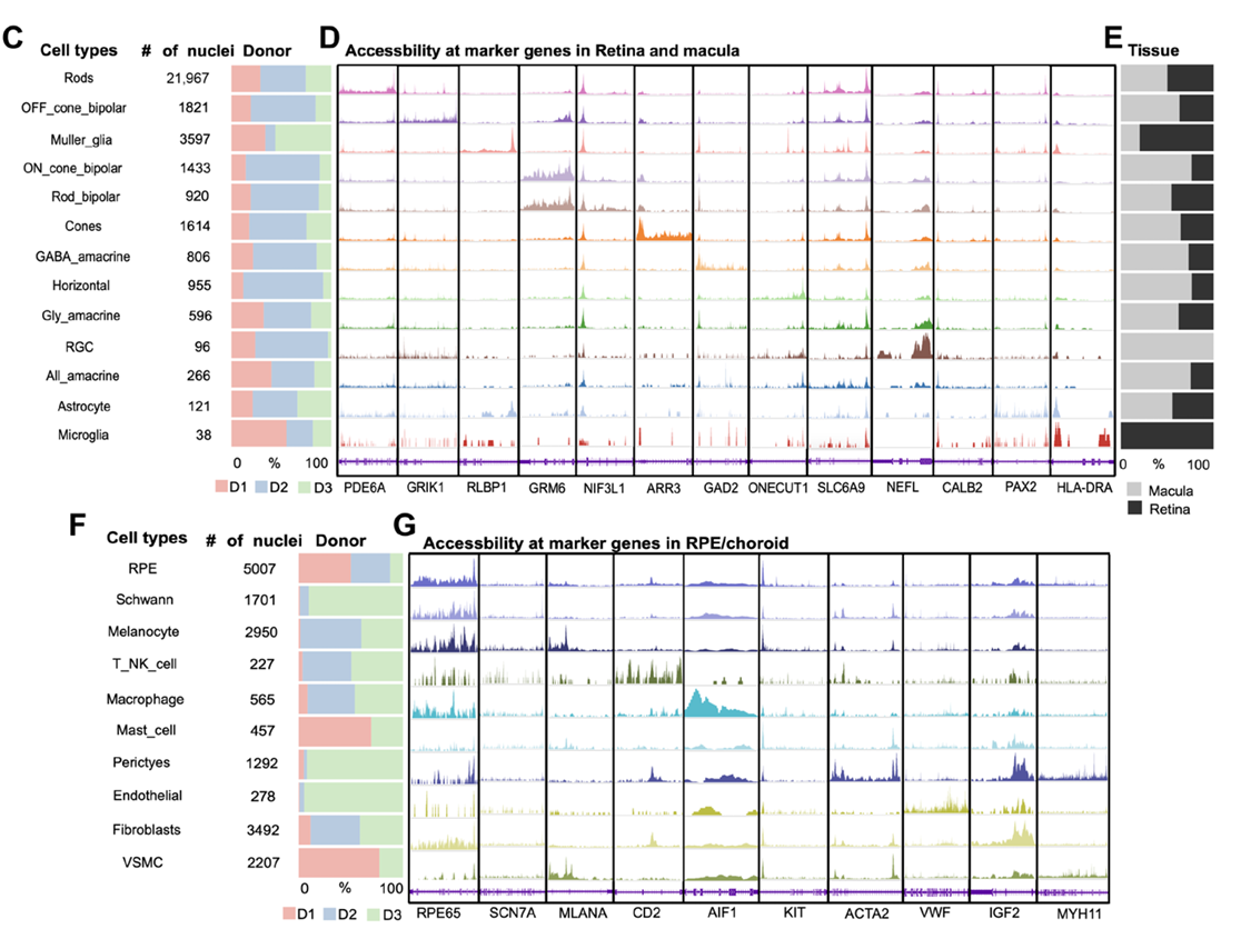
Applying single-nucleus multiome (snATAC-seq/snRNA-seq) and single-nucleus methyl-3C sequencing (snm3C-seq) to profile gene expression, chromatin accessibility, DNA methylation, and chromatin conformation helped identify over 420,000 cCREs across 23 retinal cell types, most of which displayed highly cell-type–specific accessibility patterns. An exploration of potential regulators of these cCREs revealed a comprehensive list of transcription factors, which provided a resource for a deeper understanding of the gene-regulatory networks active in human retinal cells. These data may also guide future investigations into the regulatory elements driving retinal cell-type-specific function and their implications for disease.
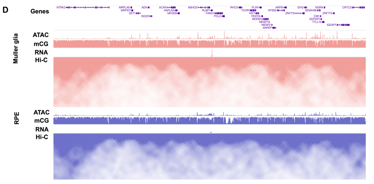
Analyzing DNA methylation data enabled the creation of a single-cell atlas of the human retina, depicting cell-type-specific DNA methylation patterns and offering insights into the regulatory landscape of each retinal cell type. Analysis of differentially methylated regions revealed over 100,000 unique cCRE-associated elements across retinal cells that typically displayed an anti-correlation pattern with cCREs. The team also explored cell-type-specific 3D genomic architecture and regulatory mechanisms; their data included the identification of ~2,000 variable topologically associating domain boundaries across retinal cells, providing insight into cell-type-specific regulatory landscapes and emphasizing cell-type-specific 3D genome organization.
The study next aimed to link cCREs to their target genes by integrating chromatin accessibility and chromatin contact maps across retinal cells; these data provided a comprehensive map of cCRE-gene interactions and highlighted the regulatory networks driving cell-specific gene expression. Notably, the authors observed varied correlation patterns, suggesting that multiple regulatory factors contribute to gene expression and underscoring the need for further investigations into the complex mechanisms that underpin retinal cell-type-specific gene regulation.
The authors then moved from human samples to mice to explore the evolutionary dynamics of cCREs and also assess the suitability of mouse models for research on human retinal diseases. A comparison of mouse and human datasets revealed consistent proportions of conserved, divergent, and human-specific cCREs across retinal cells, providing insight into the evolutionary dynamics of chromatin accessibility in the retina. Additional highlights included: i) an association of human divergent cCREs with highly expressed genes in humans; ii) the identification of epigenetic differences that reflect adaptation to unique visual demands via chromatin accessibility divergence; and iii) the conservation of transcription factor motifs, which supports the use of mouse models to explore human retinal pathological mechanisms and strengthens their relevance in translational research.
They next integrated the human retinal cell datasets with information on genetic variants associated with numerous ocular diseases; encouragingly, this analysis revealed associations between distinct diseases, cell-type-specific open chromatin profiles, and, in turn, specific cell types. For example, the team revealed that macular telangiectasia-related risk variants resided in cCREs that drive gene regulation in Müller glia, and that age-related macular degeneration-related risk variants influence gene regulatory programs specific to retinal pigment epithelium.
)
Finally, the authors trained a deep neural network to predict chromatin accessibility from DNA sequences to investigate how risk variants affect regulatory-element function in human retinal cells. In silico mutagenesis of cCREs associated with retinal disease risk variants assessed their effects on chromatin accessibility across different scenarios, revealing nuanced regulatory effects of causal and non-causal risk variants on chromatin states. The study then used CRISPR-mediated genome editing in retinal cells to validate these predictions; encouragingly, the results supported the utility of this deep-learning model in predicting the functional consequences of non-coding risk variants.
Study Strengths and How Increased Sample Size and Spatial Transcriptomics May Provide Additional Insight
The authors concluded by highlighting the strengths of their approach, which included the minimization of the impact of post-mortem intervals by using fresh retinal tissues, a deep cross-species comparison, and the exploration of the genetic underpinnings of several eye diseases. Furthermore, these data provided a more holistic view of retinal biology and its regulatory mechanisms, cell-type–specific data (including values for CREs, genes, transcription factor motifs, and CRE-gene pairs), and the first report of cell-type–specific DNA single-cell methylation data in the human retina. As limitations, the team notes the small sample size and the need to integrate single-cell multiomics with spatial transcriptomics to identify rare cell types and elucidate complex gene-regulatory networks.
Implementing Paired-Tag technology from Epigenome Technologies, which generates joint epigenetic and transcriptomic profiles at single-cell resolution and detects histone modifications and RNA transcripts in individual nuclei with efficiency comparable to single-nucleus RNA-seq/ChIP-seq assays, could provide deeper insight into these research aims. Could the simultaneous single-cell analysis of histone modifications and transcriptomic profiles have contributed to an even deeper understanding of the epigenetic and transcriptomic profiles that define the cells of the human retina?