Replication Stress Induces Genome Organization via EHMT2 and CTCF
A study reveals how links between chromatin changes and higher-order genome organization help to maintain genome integrity in response to replication stress.
Replication Stress: Do Local Chromatin Changes Impact Higher-Order Genome Organization?
The endogenous and exogenous insults that induce replication stress - a driver of genome instability and cancer (da Costa et al. and Cybulla & Vindigni) - represent a frequent challenge to DNA replication by impeding the progression of the replication fork. The protective responses employed by cells when facing such insults (Bayona-Feliu & Aguilera) include the activation of checkpoint pathways and the subsequent stabilization of stalled replication forks. During this time, the nascent DNA present at these sites remains highly vulnerable to degradation (Schlacher et al. and Berti et al.), and chromatin undergoes dynamic reorganization (González-Acosta & Lopes and Wootton & Soutoglou).
Researchers led by Nitika Taneja (Erasmus MC Cancer Institute/Oncode Institute) previously reported that the histone methyltransferase G9a promoted the de novo deposition of the repressive H3K9 methylation at stressed replication forks and local chromatin compaction, thereby protecting nascent DNA and facilitating replication fork restart (Gaggioli et al.). Their more recent Nature study sought to go one step further and explore the impact of local chromatin changes on higher-order genome organization; now, the team reports that replication stress induces anchoring of the transcriptional repressor CTCF and G9a-mediated heterochromatin assembly, which supports the formation of transient chromatin loops at stressed forks that create a protective architectural scaffold to limit nuclease access and stabilize replication intermediates (Gaggioli and Sengupta et al.)
Paired-Tag technology from Epigenome Technologies generates joint epigenetic and transcriptomic profiles at single-cell resolution and detects histone modifications and RNA transcripts in nuclei with efficiency comparable to single-nucleus RNA-seq/ChIP-seq assays. Could an in-depth analysis of the histone modification and transcriptomic profiles of the same single mesothelioma cells afforded by the integration of Paired-Tag have provided more profound insight into the impact of stress on replication forks and how the interplay between histone modifications and chromatin conformation helps to maintain genome stability?
Replication Stress Induces the Formation of G9a/CTCF-stabilized Chromatin Loops That Protect Nascent DNA
The authors initially employed their previously developed single-molecule chromatin fiber ChromStretch assay (Gaggioli et al.) to first confirm the G9a-dependent de novo deposition of H3K9me3 on nascent DNA in response to replication stress (in this case, induced by hydroxyurea which stalls replication forks). Furthermore, they developed a novel chromatin immunocleavage technique (Rep-ChIC) to systematically map H3K9me3 at active replication sites, which revealed a similar level of H3K9me3 accumulation at both early- and late-replicating regions after stress induction and indicated that replication-coupled de novo heterochromatin formation occurred globally and independently of replication timing.
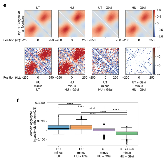
Proximity ligation assays targeting H3K9me3 at replication sites next provided evidence that G9a-mediated H3K9me3 accumulation at stressed replication sites promoted clustering of replication factories (regions rich in replication forks), suggesting that de novo heterochromatin formation at stalled forks could drive global chromosomal reorganization. Therefore, the authors performed in situ Hi-C assays and a newly developed technique called Rep-Hi-C, which enriches for interactions at newly replicated DNA, to demonstrate that G9a-mediated heterochromatin formation stabilizes replication-associated chromatin loops, thereby enhancing sister-fork interactions and establishing a replication-stress-specific genome architecture that may function to protect the genome.
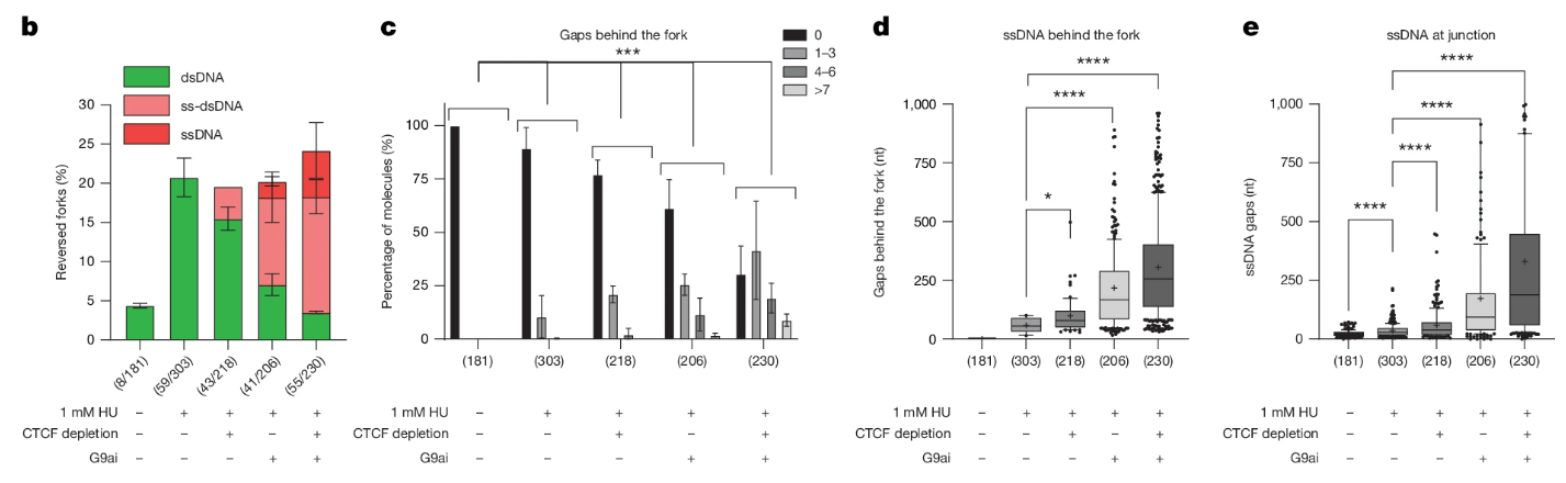
Further analysis using a wide range of cutting-edge techniques - including chromosome conformation capture coupled with quantitative PCR (3C–qPCR), DNA fluorescence in situ hybridization (DNA-FISH), TrAEL-seq (which detects fork pausing and reversal), single-cell EdU-seq, DNA fiber analysis, and a newly developed fork degradation sequencing (Fork-deg-seq) technique – together revealed that replication stress promoted CTCF-associated chromatin loop formation, which functioned to organize stressed replicons without altering the architecture of topologically associating domains at a global scale. Indeed, the authors revealed that replication stress induced the formation of G9a/CTCF-stabilized chromatin loops that marked replication fork pausing and reversal, forming a protective scaffold that could shield stalled replication forks and protect nascent DNA from degradation. In support of these findings, the study also revealed that the combined loss of stress-induced heterochromatin formation and CTCF enrichment destabilized these loop scaffolds, exposing associated DNA to nuclease activity and prompting degradation.
Maintain Genome Integrity via Chromatin Changes and Higher-Order Genome Organization
In summary, these exciting findings suggest that the local chromatin changes induced by replication stress associate with alterations in higher-order genome organization, which preserve replication fork stability and thereby mitigate mutagenesis and genomic instability by protecting the associated nascent DNA. Replication stress induces de novo H3K9me3 on nascent DNA, coinciding with the stabilization of chromatin loops that enclose stress-impacted areas; subsequently, chromatin loops become reinforced by CTCF and coated by G9a-mediated heterochromatin, which forms a protective scaffold that enhances fork stability; overall, these data provide a framework for understanding genome instability during processes such as cancer and aging.
The implementation of Paired-Tag technology from Epigenome Technologies, which generates joint epigenetic and transcriptomic profiles at single-cell resolution and detects histone modifications and RNA transcripts in individual nuclei with efficiency comparable to single-nucleus RNA-seq/ChIP-seq assays, has the potential to provide deeper insight into such research aims. Could the simultaneous single-cell analysis of histone modifications and transcriptomic profiles have contributed to the plethora of innovative techniques employed and added further depth to this fascinating study?