Can Single-Cell Transcriptomic and Epigenetic Analyses Help Develop More Faithful Brain Organoids?
.
Brain Organoids: Useful Models with Significant Room for Improvement?
Cell dysfunction in the midbrain and hindbrain, which coordinate motor movement, process sensory input, and integrate cognitive function, has been linked to the development of a range of neurological disorders (Doherty et al.). Analyses using region-specific three-dimensional brain organoids - model systems developed from human embryonic stem cells or induced pluripotent stem cells - have provided deep insight into the mechanisms underlying normal human brain development (Lancaster et al.) and the pathophysiology of neurodevelopmental disorders (Li et al.). Of note, recent single-cell transcriptomic analyses of brain organoids have highlighted the underrepresentation of specific neuron populations (He et al.), underscoring our inability to create organoids that accurately reflect specific brain regions (Fiorenzano et al.). This limitation stems from a lack of information regarding the optimized morphogen timings, concentrations, and combinations required during organoid development, and on how they converge on transcriptional regulation to guide the specification of regional cell types. How can we develop more faithful brain organoid models containing underrepresented cell types?
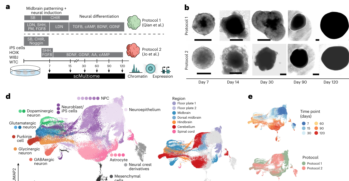
To understand more regarding current midbrain/hindbrain organoid models and explore potential improvements to the protocols employed for their formation, researchers led by J. Gray Camp (Roche Innovation Center Basel/University of Basel) and Barbara Treutlein (ETH Zürich) recently used paired single-cell transcriptome and accessible chromatin sequencing to map out cell compositions and define regulatory mechanisms and also morphogen screening to identify the specific conditions that could drive the emergence of underrepresented cell types. Overall, their new Nature Neuroscience study provides deeper insight into human midbrain/hindbrain organoid development by defining cellular complexity through joint transcriptomic and epigenetic analyses and reports new protocols that support the development of critically important region-specific cell types and, as such, more faithful brain organoid models. (Azbukina, He, and Lin et al.).
Paired-Tag technology from Epigenome Technologies generates joint epigenetic and transcriptomic profiles at single-cell resolution and detects histone modifications and RNA transcripts in nuclei with efficiency comparable to single-nucleus RNA-seq/ChIP-seq assays. Could an in-depth analysis of histone modifications and transcriptomic profiles of the same single cells afforded by the integration of Paired-Tag have provided complementary data and allowed an even deeper understanding of the composition of midbrain/hindbrain organoids and improvements to associated protocols to generate more faithful brain organoid models?
Can Single-Cell Transcriptomic and Epigenetic Analyses Improve Protocols for Brain Organoid Generation?
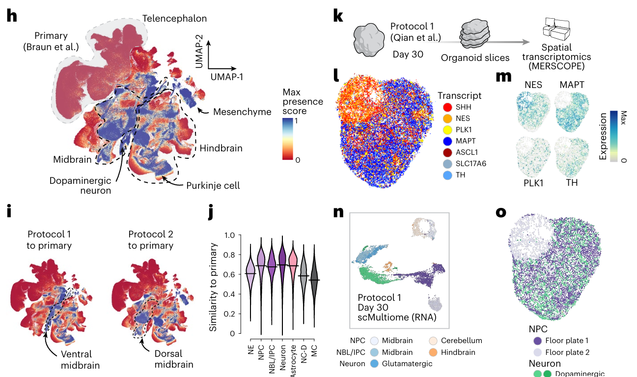
The authors applied simultaneous single-cell transcriptomic and chromatin accessibility profiling to brain organoids developed using two existing protocols originally employed to generate midbrain cell types (Qian et al. and Jo et al.), which revealed the development of ventral and dorsal cell types that cover regions such as the floor plate, dorsal and ventral midbrain, and adjacent hindbrain regions. Of note, this approach goes beyond simply evaluating gene transcription data and provides insight into the regulatory landscape of these complex brain regions. The fact that these protocols generated significant numbers of hindbrain cells indicated a need to improve in vitro culture protocols to achieve greater regional specificity; however, these data also underscore the importance of single-cell profiling of organoids for benchmarking and validating cell-type diversity. The team then applied gene regulatory network inference and transcription factor perturbation via CRISPR to gain deeper insights into the mechanisms underlying human brain regionalization/neuronal differentiation; overall, they identified brain-region-specific programs involving well-known master regulators, including transcription factors such as OTX2, LMX1A, LHX1, EN2, and ZIC3. In addition, they found evidence that similar genetic programs helped establish and maintain regional identity, providing support for a deterministic specification of neural diversity (Ge et al.).
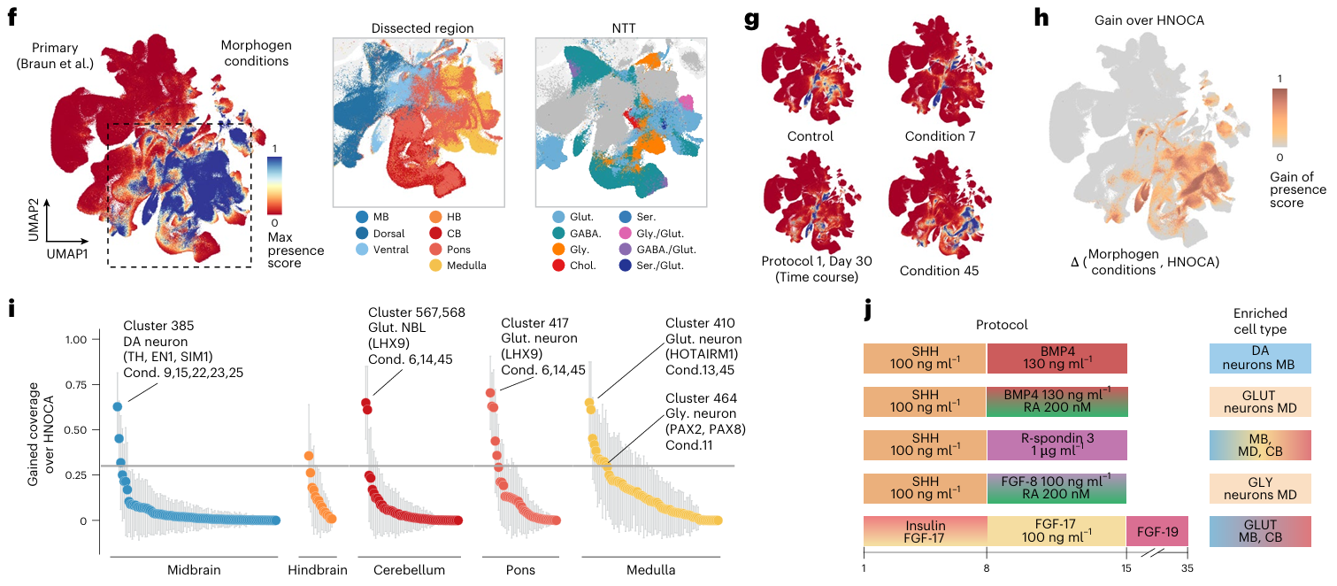
The study then employed a single-cell multiplexed patterning screen to expand and optimize protocols for brain organoid formation, thereby increasing the diversity of cell types. Excitingly, this approach identified specific morphogen concentrations and morphogen combinations that promoted the development of underrepresented brain cell types, including protocols that yielded cerebellum-specific and pons-specific glutamatergic neurons, glutamatergic neurons of the dorsal medulla, and glycinergic neurons of the ventral medulla. Importantly, the authors note the potential applications of organoid models containing these cells, which included: i) the advanced modeling of hyperekplexia-like disorders (characterized by pronounced startle responses to tactile or acoustic stimuli and ensuing periods of spasticity/rigidity; Safory et al., Zafra et al., and Davies et al.) when considering glycinergic neurons; ii) the study of mechanisms of autism spectrum disorder development (Pilorge et al., Nisar et al., and McKimm et al.) when considering glycinergic neurons and cerebellar glutamatergic neurons; and iii) the evaluation of targeted therapeutics for known driver genes and the exploration of novel, currently unknown driver genes in pediatric brain tumors.
To conclude, the authors noted certain limitations of their exciting study, which they aim to address in the future by applying multiplexed protein staining or functional assays to provide further biological insights and complement their highly interesting findings.
Single-Cell Transcriptomic and Epigenetic Analyses: The Way Forward for Improved Brain Organoids?
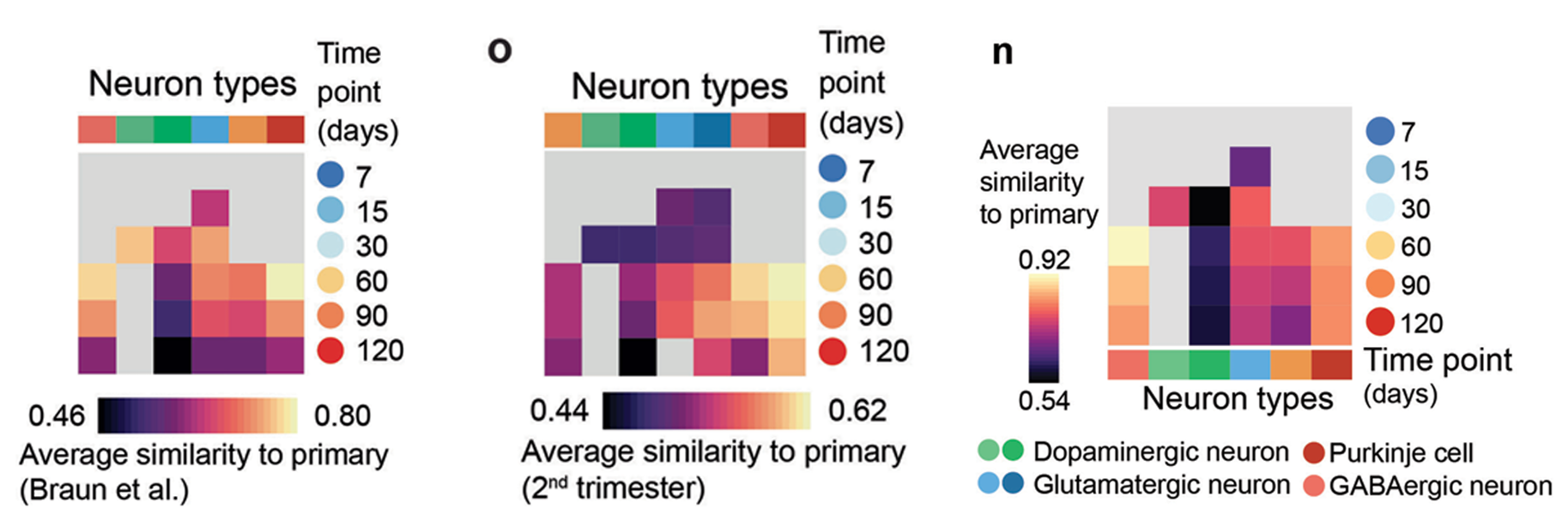
Overall, the application of single-cell transcriptomic and epigenetic analyses has advanced our understanding of the complexity and developmental dynamics of human midbrain/hindbrain organoid development and has improved protocols for creating more faithful brain organoid models. The implementation of Paired-Tag technology from Epigenome Technologies, which generates joint epigenetic and transcriptomic profiles at single-cell resolution and detects histone modifications and RNA transcripts in individual nuclei with efficiency comparable to single-nucleus RNA-seq/ChIP-seq assays, has the potential to provide deeper insight into such research aims. Could the simultaneous single-cell analysis of histone modification and transcriptomic profiles have provided complementary data that would have revealed more regarding our understanding of the composition of midbrain/hindbrain organoids and supported additional improvements to associated protocols?