Single-Cell Chromatin and Transcriptomic Analyses of Tauopathy-affected Brains Reveal How Glial Cells Contribute
A study employing single-cell chromatin accessibility and transcriptomic analyses of brain samples from tauopathy individuals reveals the contribution of glia.
Exploring the Contribution of Glial Cells to Neurodegenerative Tauopathy
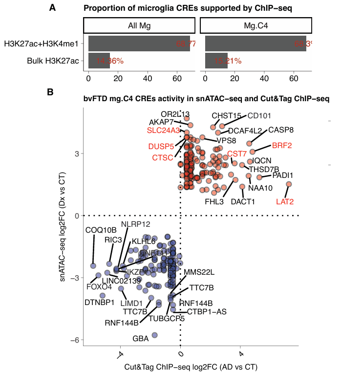
Recent research from the laboratory of Jessica E. Rexach and Daniel H. Geschwind (University of California, Los Angeles) revealed the diversity of shared and distinct cellular responses to tau-associated disorders - Pick's disease (PiD), progressive supranuclear palsy (PSP), and Alzheimer's disease (AD) - that affect distinct neuron and glial cell subpopulations within disorder-specific brain regions (Rexach et al., 2020 and Rexach et al., 2024). Abnormal tau aggregation in the brain characterizes these neurodegenerative "tauopathies," whose symptoms include dementia and Parkinsonism (Moloney et al., Debnath et al., and Chung et al.). Specifically, their studies described an AD-enriched microglia state characterized by the elevated expression of genes associated with AD risk and those known to protect against AD-specific pathology. How can we further understand the tauopathy-affected brain?
Their subsequent research aimed to explore whether glial cell subpopulations possess distinct mechanisms underlying their diverse roles in the pathogenesis of these tauopathies, and to determine how chromatin remodeling integrates genetic risk and regulatory variation across tauopathies and affected brain regions. Now, a new study from the Jessica E. Rexach laboratory reports on the application of single-nucleus profiling of chromatin accessibility and gene expression across AD, PiD, and PSP in brain regions displaying distinct vulnerabilities (calcarine of the visual cortex, insular cortex, and precentral gyrus of the frontal cortex) to comprehensively investigate epigenetic dysregulation in glial cell subpopulations. Here, we provide a summary of the major biological discoveries, translational implications, limitations, and future directions described in this recently published Nature Communications article (Han et al.).
Paired-Tag technology from Epigenome Technologies generates joint epigenetic and transcriptomic profiles at single-cell resolution and detects histone modifications and RNA transcripts in nuclei with efficiency comparable to single-nucleus RNA-seq/ChIP-seq assays. Could an in-depth analysis of histone modifications and transcriptomic profiles of the same single cells via the integration of Paired-Tag reveal more regarding the contribution of glial cells to the development of taupothies?
Single-Cell Chromatin and Transcriptomic Analyses Reveal the Glial Contribution to Tauopathy
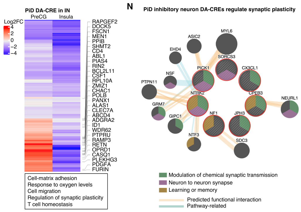
This exciting study reported analyses of chromatin accessibility (snATAC-seq) and transcriptomic (snRNA-seq) profiles from >600,000 nuclei isolated from brain regions of 41 individuals with tauopathies. This approach permitted the identification of both cell-type- and context-specific cis-regulatory elements (CREs) - regions of non-coding DNA that regulate the transcription of neighboring genes - and the characterization of changes in chromatin accessibility. The authors identified regions that displayed disease-associated dynamism and interrogated genetic variants from genome-wide association studies (GWAS), single-nucleus expression quantitative trait loci (eQTLs), and functional variants validated using massively parallel reporter assays (MPRAs) at these regions. This strategy helped in the definition of critical regulatory transcription factors, the prioritization of target genes, and the identification of non-coding regulatory circuits disrupted in specific disease-affected cells. Overall, this permitted the authors to define disease-associated glial states - PiD-related microglia and PSP-related astrocytes - that undergo expansion in the disease-affected insular cortex and display disease-linked and coordinated epigenomic, transcriptional, and functional signatures.
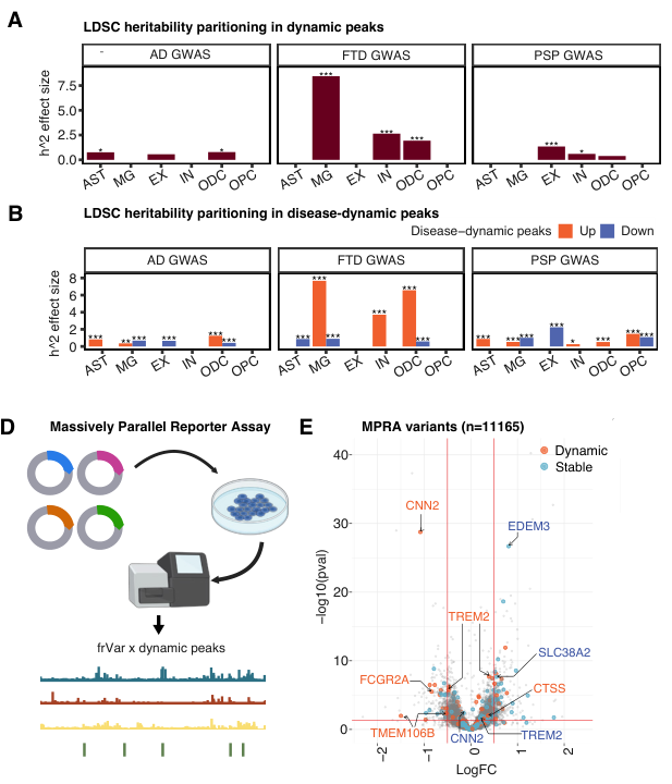
The data from this study highlighted that chromatin accessibility dynamics effectively captured disease heritability and functional genetic variation; overall, the findings underscored a conserved transition in chromatin accessibility when shifting from moderately to severely affected brain regions, reflecting a coordinated program of regulatory activation and repression during disease progression. The authors also revealed that CREs displaying disease-associated dynamism contained a disproportionate number of GWAS variants, single-nucleus eQTLs, and MPRA-validated functional variants. Analysis of these CREs also helped identify transcription factor-centered regulatory modules; for example, the transcription factors MEF2C and MEF2D formed a regulatory hub that linked genetic variation to lysosomal, phagocytic, and stress-responsive programs in disease-associated microglia.
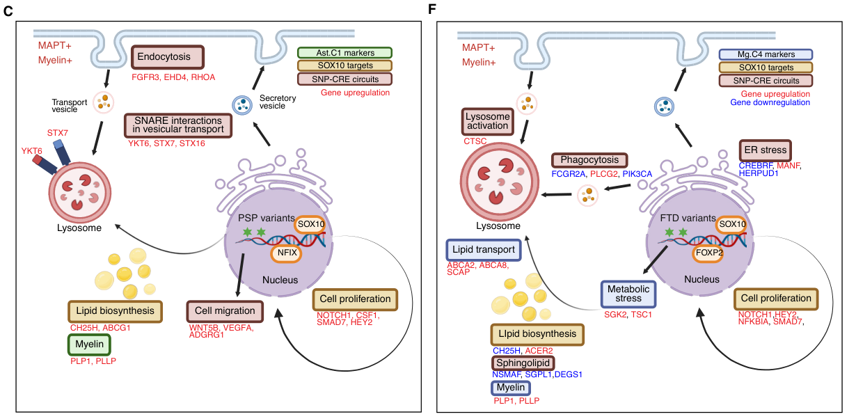
When considering glial expansion as a conserved response to neurodegeneration, the authors found that PiD-related microglial expansion concentrated disease heritability in CREs that control lipid and debris clearance programs, while PSP-related astrocytic expansion engaged SNARE-dependent trafficking pathways by preserving chromatin accessibility and risk-variant-linked enhancers. Together, these data suggest that disease-specific genetic risk shapes glial resilience by selectively tuning lysosome-vesicle pathways in distinct cell types.
The Glial Contribution to Tauopathy: Translational Implications, Limitations, and Future Directions
Overall, combined epigenetic and transcriptomic analysis provided evidence that common genetic risk factors act through disease-context-specific CREs to modulate glial responses rather than uniformly activating inflammatory pathways. The data also revealed candidate genes and pathways selectively engaged in disease-relevant glial states, including lysosomal trafficking, sphingolipid metabolism, vesicle fusion, and phagocytosis, and identified regulatory circuits that may represent therapeutic targets to enhance glial resilience. However, the authors note limitations in their study, including the need to integrate proteomic and spatial readouts and to move evaluations beyond end-stage human brain tissue. Their future research aims include perturbing CREs and transcription factors in disease-relevant models to evaluate their impact on cellular function and to include additional neurodegenerative disorders.
The implementation of Paired-Tag technology from Epigenome Technologies, which generates joint epigenetic and transcriptomic profiles at single-cell resolution and detects histone modifications and RNA transcripts in individual nuclei with efficiency comparable to single-nucleus RNA-seq/ChIP-seq assays, has the potential to provide deeper insight into such research aims. Could the simultaneous single-cell analysis of histone modification and transcriptomic profiles help to define how glial cells contribute to the development of human tauopathies? Of note, the authors thank Epigenome Technologies' very own Christopher Hartl for sharing CUT&Tag data, which contributed to the development of this exciting new study.